2-Minute Neuroscience: Electroencephalography (EEG)
Electroencephalography, or EEG, is a technique used to measure the electrical activity of the brain. In this video, I discuss the basics of EEG like what it measures, how it’s used, and some strengths and limitations.
TRANSCRIPT:
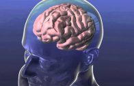
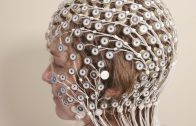
Electroencephalography, or EEG, is a technique used to measure the electrical activity of the brain. In EEG, electrodes are most commonly placed on the scalp of a patient, primarily to detect the electrical activity of neurons in the cerebral cortex. Typically, EEG does not record the activity of single neurons, but rather detects the signals created when populations of neurons are active at the same time. It mostly records signals from small areas of the brain surrounding each electrode. EEG is primarily measuring postsynaptic potentials, or changes in membrane potential that are elicited by neurotransmitters binding to receptors on the postsynaptic membrane.
EEG provides an image of electrical activity in the brain represented as waves of varying frequency, amplitude, and shape. It can be used to measure brain activity that occurs during an event–like the completion of a task or the presentation of a stimulus–or to measure spontaneous brain activity that happens in the absence of a specific event. The typical electrical activity that occurs in association with an event is sometimes referred to as the event-related potential. There are a number of clinical applications of EEG. For example, it is commonly used to diagnose epilepsy and characterize seizure activity, to monitor sleep for the diagnosis of sleep disorders, and in efforts to diagnose or provide information about a variety of other types of brain dysfunction.
While there are many advantages to using EEG, such as its low cost and ability to measure brain activity on the order of milliseconds, there are some limitations as well. For example, because EEG monitors activity in large groups of neurons, it is difficult to pinpoint activity seen using EEG to a precise location in the brain. And, it records the activity of neurons in the cortex, but is limited in its ability to accurately record activity in deeper structures of the brain.
REFERENCES:
Biasiucci A, Franceschiello B, Murray MM. Electroencephalography. Curr Biol. 2019 Feb 4;29(3):R80-R85. doi: 10.1016/j.cub.2018.11.052.
Frey LC, Spitz MC. 2013. Electroencephalography. In: Arciniegas DB, Anderson CA, Filley CM, eds. Behavioral Neurology & Neuropsychiatry. New York: Cambridge University Press.
The image of EEG output used in this video is a CC image courtesy of Andrii Cherninskyi and was downloaded from: https://commons.wikimedia.org/wiki/File:Human_EEG_with_prominent_alpha-rhythm.png